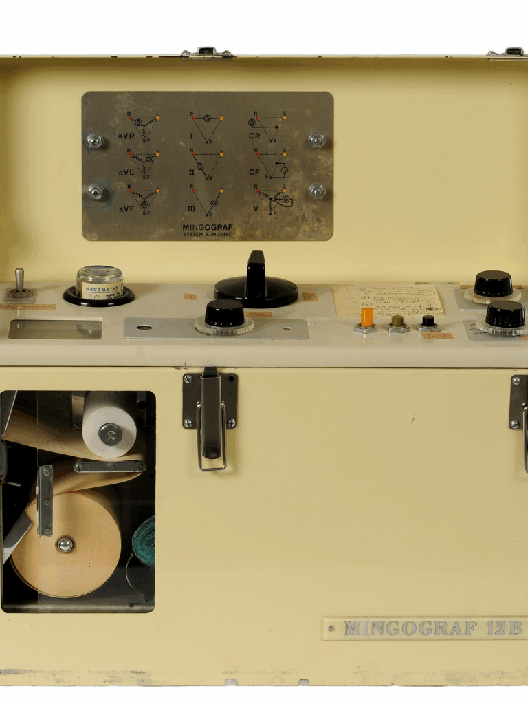
Millions of people worldwide have reason to be thankful that Swedish engineer Rune Elmqvist decided not to practice medicine. Although qualified as a doctor, he chose to invent medical equipment instead. In 1949, while working at Elema-Schonander (later Siemens-Elema), in Stockholm, he applied for a patent for the Mingograph, the first inkjet printer. Its movable nozzle deposited an electrostatically controlled jet of ink droplets on a spool of paper.
 Rune Elmqvist qualified to be a physician, but he devoted his career to developing medical equipment, like this galvanometer.Håkan Elmqvist/Wikipedia
Elmqvist demonstrated the Mingograph at the First International Congress of Cardiology in Paris in 1950. It could record physiological signals from a patient’s electrocardiogram or electroencephalogram in real time, aiding doctors in diagnosing heart and brain conditions. Eight years later, he worked with cardiac surgeon Åke Senning to develop the first fully implantable pacemaker. So whether you’re running documents through an inkjet printer or living your best life due to a pacemaker, give a nod of appreciation to the inventive Dr. Elmqvist.
The world’s first inkjet printer
Rune Elmqvist was an inquisitive person. While still a student, he invented a specialized potentiometer to measure pH and a portable multichannel electrocardiograph. In 1940, he became head of development at the Swedish medical electronics company Elema-Schonander.
Before the Mingograph, electrocardiograph machines relied on a writing stylus to trace the waveform on a moving roll of paper. But friction between the stylus and the paper prevented small changes in the electrical signal from being accurately recorded. Elmqvist’s initial design was a modified oscillograph. Traditionally, an oscillograph used a mirror to reflect a beam of light (converted from the electrical signal) onto photographic film or paper. Elmqvist swapped out the mirror for a small, moveable glass nozzle that continuously sprayed a thin stream of liquid onto a spool of paper. The electrical signal electrostatically controlled the jet.
 The Mingograph was originally used to record electrocardiograms of heart patients. It soon found use in many other fields.Siemens Healthineers Historical Institute
By eliminating the friction of a stylus, the Mingograph (which the company marketed as the Mingograf) was able to record more detailed changes of the heartbeat. The machine had three paper-feed speeds: 10, 25, and 50 millimeters per second. The speed could be preset or changed while in operation.
RELATED: The Inventions That Made Heart Disease Less Deadly
An analog input jack on the Mingograph could be used to take measurements from other instruments. Researchers in disciplines far afield from medicine took advantage of this input to record pressure or sound. Phoneticians used it to examine the acoustic aspects of speech, and zoologists used it to record birdsongs. Throughout the second half of the 20th century, scientists cited the Mingograph in their research papers as an instrument for their experiments.
Today, the Mingograph isn’t that widely known, but the underlying technology, inkjet printing, is ubiquitous. Inkjets dominate the home printer market, and specialized printers print DNA microarrays in labs for genomics research, create electrical traces for printed circuit boards, and much more, as Phillip W. Barth and Leslie A. Field describe in their 2024 IEEE Spectrum article “Inkjets Are for More Than Just Printing.”
The world’s first implantable pacemaker
Despite the influence of the Mingograph on the evolution of printing, it is arguably not Elmqvist’s most important innovation. The Mingograph helped doctors diagnose heart conditions, but it couldn’t save a patient’s life by itself. One of Elmqvist’s other inventions could and did: the first fully implantable, rechargeable pacemaker.
 The first implantable pacemaker [left] from 1958 had batteries that needed to be recharged once a week. The 1983 pacemaker [right] was programmable, and its batteries lasted several years.Siemens Healthineers Historical Institute
Like many stories in the history of technology, this one was pushed into fruition at the urging of a woman, in this case Else-Marie Larsson. Else-Marie’s 43-year-old husband, Arne, suffered from scarring of his heart tissue due to a viral infection. His heart beat so slowly that he constantly lost consciousness, a condition known as Stokes-Adams syndrome. Else-Marie refused to accept his death sentence and searched for an alternative. After reading a newspaper article about an experimental implantable pacemaker being developed by Elmqvist and Senning at the Karolinska Hospital in Stockholm, she decided that her husband would be the perfect candidate to test it out, even though it had been tried only on animals up until that point.
External pacemakers—that is, devices outside the body that regulated the heart beat by applying electricity—already existed, but they were heavy, bulky, and uncomfortable. One early model plugged directly into a wall socket, so the user risked electric shock.
By comparison, Elmqvist’s pacemaker was small enough to be implanted in the body and posed no shock risk. Fully encased in an epoxy resin, the disk-shaped device had a diameter of 55 mm and a thickness of 16 mm—the dimensions of the Kiwi Shoe Polish tin in which Elmqvist molded the first prototypes. It used silicon transistors to pace a pulse with an amplitude of 2 volts and duration of 1.5 milliseconds, at a rate of 70 to 80 beats per minute (the average adult heart rate).
The pacemaker ran on two rechargeable 60-milliampere-hour nickel-cadmium batteries arranged in series. A silicon diode connected the batteries to a coil antenna. A 150-kilohertz radio loop antenna outside the body charged the batteries inductively through the skin. The charge lasted about a week, but it took 12 hours to recharge. Imagine having to stay put that long.
 In 1958, over 30 years before this photo, Arne Larsson [right] received the first implantable pacemaker, developed by Rune Elmqvist [left] at Siemens-Elema. Åke Senning [center] performed the surgery.Sjöberg Bildbyrå/ullstein bild/Getty Images
Else-Marie’s persuasion and persistence pushed Elmqvist and Senning to move from animal tests to human trials, with Arne as their first case study. During a secret operation on 8 October 1958, Senning placed the pacemaker in Arne’s abdomen wall with two leads implanted in the myocardium, a layer of muscle in the wall of the heart. The device lasted only a few hours. But its replacement, which happened to be the only spare at the time, worked perfectly for six weeks and then off and on for several more years.
 Arne Larsson lived another 43 years after his first pacemaker was implanted. Shown here are five of the pacemakers he received. Sjöberg Bildbyrå/ullstein bild/Getty Images
Arne Larsson clearly was happy with the improvement the pacemaker made to his quality of life because he endured 25 more operations over his lifetime to replace each failing pacemaker with a new, improved iteration. He managed to outlive both Elmqvist and Senning, finally dying at the age of 86 on 28 December 2001. Thanks to the technological intervention of his numerous pacemakers, his heart never gave out. His cause of death was skin cancer.
Today, more than a million people worldwide have pacemakers implanted each year, and an implanted device can last up to 15 years before needing to be replaced. (Some pacemakers in the 1980s used nuclear batteries, which could last even longer, but the radioactive material was problematic. See “The Unlikely Revival of Nuclear Batteries.”) Additionally, some pacemakers also incorporate a defibrillator to shock the heart back to a normal rhythm when it gets too far out of sync. This lifesaving device certainly has come a long way from its humble start in a shoe polish tin.
Rune Elmqvist’s legacy
Whenever I start researching the object of the month for Past Forward, I never know where the story will take me or how it might hit home. My dad lived with congestive heart failure for more than two decades and absolutely loved his pacemaker. He had a great relationship with his technician, Francois, and they worked together to fine-tune the device and maximize its benefits. And just like Arne Larsson, my dad died from an unrelated cause.
An engineer to the core, he would have delighted in learning about the history of this fantastic invention. And he probably would have been tickled by the fact that the same person also invented the inkjet printer. My dad was not a fan of inkjets, but I’m sure he would have greatly admired Rune Elmqvist, who saw problems that needed solving and came up with elegantly engineered solutions.
Part of a continuing series looking at historical artifacts that embrace the boundless potential of technology.
An abridged version of this article appears in the September 2025 print issue.
References
There is frustratingly little documented information about the Mingograph’s origin story or functionality other than its patent. I pieced together how it worked by reading the methodology sections of various scientific papers, such as Alf Nachemson’s 1960 article in Acta Orthopaedica Scandinavica, “Lumbar Intradiscal Pressure: Experimental Studies on Post-mortem Material”; Ingemar Hjorth’s 1970 article in the Journal of Theoretical Biology, “A Comment on Graphic Displays of Bird Sounds and Analyses With a New Device, the Melograph Mona”; and Paroo Nihalani’s 1975 article in Phonetica, “Velopharyngeal Opening in the Formation of Voiced Stops in Sindhi.” Such sources reveal how this early inkjet printer moved from cardiology into other fields.
Descriptions of Elmqvist’s pacemaker were much easier to find, with Mark Nicholls’s 2007 profile “Pioneers of Cardiology: Rune Elmqvist, M.D.,” in Circulation: Journal of the American Heart Association, being the main source. Siemens also pays tribute to the pacemaker on its website; see, for example, “A Lifesaver in a Plastic Cup.”