When Xiangyi Cheng published her first journal paper as a principal investigator in IEEE Access in 2024, it marked more than a professional milestone. For Cheng, an IEEE member and an assistant professor of mechanical engineering at Loyola Marymount University, in Los Angeles, it was the latest waypoint in a career shaped by curiosity, persistence, and a belief that technology should serve people—not the other way around.
The paper’s title was “Mobile Devices or Head-Mounted Displays: A Comparative Review and Analysis of Augmented Reality in Healthcare.”
XIANGYI CHENG
Employer
Loyola Marymount University, in Los Angeles
Title
Assistant professor of mechanical engineering
Member grade
Member
Alma maters
China University of Mining and Technology; Texas A&M University
Cheng’s work spans robotics, intelligent systems, human-machine interaction and artificial intelligence. It has applications in patient-specific surgical planning, an approach whereby treatment is customized to the anatomy and clinical needs of each individual.
Her research also covers wearables for rehabilitation and augmented-reality-enhanced engineering education.
The throughline of her career is sound judgment based on critical thinking. She urges her students to avoid the temptation to accept the answers they’re given by AI without cross-checking them against their own foundational understanding of the subject matter.
“AI can give you ideas,” Cheng says, “but it should never lead your thinking.”
That principle—honed through uncertainty, disciplinary shifts, and hard-earned confidence—has made Cheng an emerging voice in applied intelligent systems and a thoughtful educator preparing students for an AI-saturated world.
From Xi’an to Beijing: A mind drawn to mathematics
Cheng, born in Xi’an, China, grew up in a household shaped by her parents’ disparate careers. Her father was a mining engineer, and her mother taught Chinese and literature at a high school.
“That contrast between logical and literary thinking helped me understand myself early,” Cheng says. “I liked math, and STEM felt natural to me.”
Several teachers reinforced her inclination, she says, particularly a math teacher whose calm, fair approach emphasized reasoning over punishments such as detention for misbehavior or failure to complete assignments.
“It wasn’t about being right,” Cheng says. “It was about thinking clearly.”
She moved to Beijing in 2011 to attend the China University of Mining and Technology , where she studied mechanical engineering. After graduating with a bachelor’s degree in 2015, she was unsure where the field would take her.
An IEEE paper changed her trajectory
Later in 2015, she traveled to the United States to study at Case Western Reserve University, in Cleveland.
She initially viewed the move as exploratory rather than a long-term commitment.
“I wasn’t thinking about a Ph.D.,” she says. “I wasn’t even sure research was for me.”
That uncertainty shifted in 2017, when Cheng submitted her “IntuBot: Design and Prototyping of a Robotic Intubation Device” paper to the IEEE International Conference on Robotics and Automation (ICRA)—which was accepted.
“AI can give you more possibilities, but thinking is still our responsibility.”
Intubation is a procedure in which an endotracheal tube is inserted into a patient’s airway—usually through the mouth—to help them breathe. Because placing the tube correctly is not simple and usually must be done quickly, it requires training. That’s why research into robotic or assisted intubation systems focuses on improving speed, accuracy, and safety.
She presented her findings at ICRA in 2018, giving her early exposure to a global research community.
“That acceptance gave me confidence,” she recalls. “It showed me I could contribute to the field.”
Her advisor at Case Western encouraged her to switch from the mechanical engineering master’s program to the Ph.D. track. When the advisor moved to Texas A&M University, in College Station, in 2019, Cheng decided to transfer. She completed her Ph.D. in mechanical engineering at Texas A&M in 2022.
Although she didn’t earn a degree from Case Western, she credits her experience there with clarifying her professional direction.
Shortly after graduating with her Ph.D., Cheng was hired as an assistant professor of mechanical engineering at Ohio Northern University, in Ada. She left in 2024 to become an assistant professor at Loyola Marymount.
Engineering for the body—and the classroom
Cheng’s research focuses on human-centered engineering, particularly in health care. One of her major projects addresses syndactyly, a congenital condition in which a newborn’s fingers are fused at birth. Surgeons rely on their experience to estimate the size and shape of skin grafts to be taken from another part of the body for the corrective surgery.
She is developing technology to scan the patient’s hand, extract anatomical landmarks, and use finite element analysis—a computer-based method for predicting how a physical object will behave under real-world conditions—to determine the optimal graft size and shape.
 Xiangyi Cheng designs human-centered intelligent systems with applications in health care and education.Xiangyi Cheng
“Everyone’s hand is different,” Cheng says. “So the surgery should be personalized.”
Another project centers on developing smart gloves to assist with hand rehabilitation, pairing the unaffected hand with the injured one so the person’s natural motion can help guide therapy.
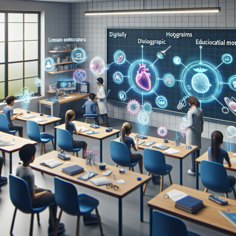
She also is exploring augmented reality in engineering education, using immersive visualization and AI tools to help students grasp three-dimensional concepts that are difficult to convey through traditional learning tools. Such visualization lets students see and interact with a digital world as if they’re inside it instead of viewing it on a flat screen.
Teaching balance in an AI-driven world
Despite working at the forefront of AI-enabled systems, Cheng cautions her students to be judicious in their use of the technology so that they don’t rely on it too heavily.
“AI is not always right and perfect,” she says. “You still need to be able to judge whether the answers it provides are correct.”
As AI continues to reshape engineering, Cheng remains grounded in a simple principle, she says: “We should use these tools. But we should never let them replace our judgment. AI can give you more possibilities, but thinking is still our responsibility.”
In her lab and classroom, Cheng prioritizes independent thinking, critical evaluation, and persistence. Many of her research students are undergraduates, and she encourages them to take ownership of their work—planning ahead, testing ideas, and learning from failure.
“The students who succeed don’t give up easily,” she says.
What she finds most rewarding, she says, is watching students mature. Reserved first-year students often become confident seniors who can present complex work and manage demanding projects.
“Getting to witness that transformation is why I teach,” she says.
For students considering engineering, Cheng offers straightforward advice: “Focus on mathematics. Engineering looks hands-on, but math is the foundation behind everything.”
With practice and persistence, she says, students can succeed and find meaning in the field.
Why IEEE continues to matter
Cheng joined IEEE in 2017, the year she submitted her first paper to ICRA. The organization has remained central to her professional development, she says.
She has served as a reviewer for IEEE journals and conferences including Robotics and Automation Letters, Transactions on Medical Robotics and Bionics, Transactions on Robotics, the International Conference on Intelligent Robots and Systems, and ICRA.
IEEE’s interdisciplinary scope aligns naturally with her work, she says, adding that the organization is “one of the few places that truly welcomes research across boundaries.”
More personally, IEEE helped her see a future she had not initially imagined.
“That first conference was a turning point,” she says. “It helped me realize I belonged.”